![]()
Art transports us, engages us, provokes thought, summons deep emotions, mirrors experience, and, most often, is simply beautiful.
Whether viewing art or creating art, the relationship between art and the brain is undeniable.
Inspired by neuroscience research at the Friedman Brian Institute, the Art of the Brain exhibition, created and curated by Veronica Szarejko, Director of the Art of the Brain, is a collection of photographs, illustrations and sculptures that celebrate the beauty of the brain as seen through the eyes of some of the world’s leading researchers and medical illustrators. With the aid of the latest technological advances, scientists are better able to understand how the brain works and to accelerate the development of new treatments for many brain disorders including Alzheimer’s disease, autism, drug addiction, schizophrenia and Parkinson’s disease, among many others. Whether an “accidental” element or an intended consequence, the byproduct of this research, the pieces that make up the Art of the Brain exhibition, offer an “explosion of vibrant colour and intricate detail, evoking chaos and calm, beauty and pain…” An experience that “is about being human; the remarkable microscopic activity that makes us living feeling beings—cognition, emotion, sensation, perception.”
Born from the Art of the Brain exhibition, the Art of the Brain Lecture series features an exciting array of experts from within the science and art communities with whom we explore the intersection of art and neuroscience and the variety of ways in which this convergence impacts people on both a neurological level and the level of human experience.
Both the Art of the Brain exhibition and the lecture series expand the Friedman Brain Institute’s reach beyond an academic/scientific setting to laypeople of all ages connecting the Friedman Brain Institute with its East Harlem community on a visceral level as they “offer many perspectives, interpretations and insights” into the human condition.
By Selene Lomoio, PhD & Rachel Willen
Where Neurons Dream
Department of Neurology
Two architectures of the same stem cell–derived human neural culture: a controlled chip below and a soft 3D silk scaffold above. Axons rise like trees toward constellations of 3D cultures, tracing the emergence of connection across engineered space. Together, it shows how these platforms can recreate the growth and linking of human neural networks.


By Natalie Suhy
Patient Stem Cell-Derived Blood-Brain Barrier In A Dish
Department of Neuroscience
3D patient stem cell-derived blood-brain barrier model created in a dish to study genetic drivers of Alzheimer’s disease pathology, stained for microvascular endothelial cells
Dina Vazquez Carrillo, PhD
Nash Family Department of Neuroscience
The Universe of the Brain
Immunofluorescent staining of the GLP-1 receptor in the septal region of the mouse brain. The receptor appears in blue, while neuronal nuclei glow in gold (DAPI), revealing a hidden stellar landscape within neural tissue.

By Madeline Cheshire
Nash Family Department of Neurology
Hecate Anatome
Three anatomical drawings of the brain, inspired by reference images from Thomas Willis’ “Cerebri Anatome” (1664) with illustrations by Sir Christopher Wren. Ink and watercolor on paper.

Melissa Umphlett, MD
Department of Pathology (Neuropathology division)
Neuron #1
Acrylic on canvas panel 11 x 14 inch (w x h)
This painting depicts a neuron and is the first in a series of paintings with the same theme.

Anurupa Ghosh, PhD
Department of Pharmacological Sciences
Mice Hippocampal Formation
Mice hippocampus staining with NeuN (yellow), Acetyle Choline (Red), vGAD (Green), DAPI (Blue)
Brian Schilder, M.Phil
Department of Genetics & Genomic Sciences
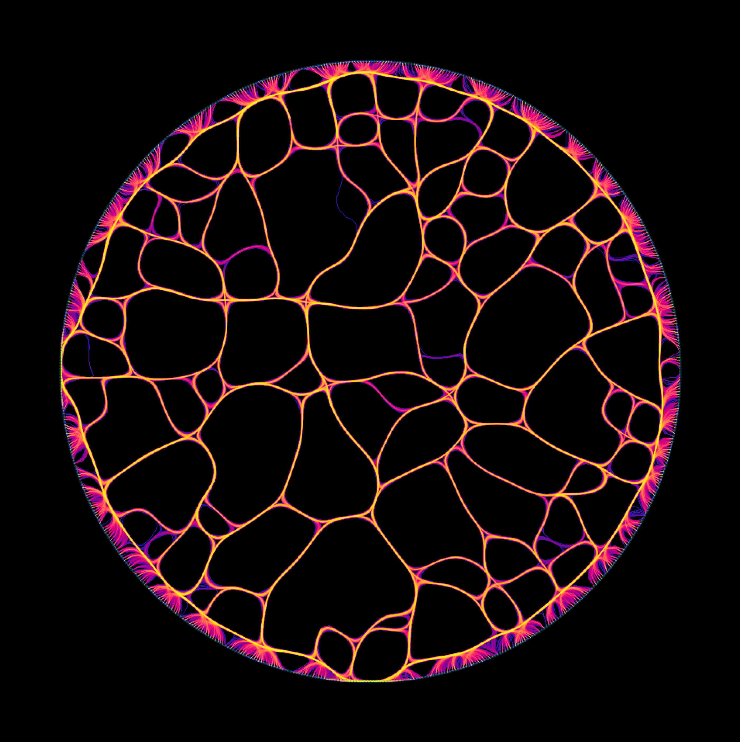
Wildfire
Transcriptomic data from 16k+ individual brain cells (shown as points) after reducing the dimensionality with an autoencoder and UMAP. 5 million tracts are shown interconnecting these cells, where shorter tract length represents greater similarity in their molecular profiles.
Jacob Wolf
Department of Neurosurgery
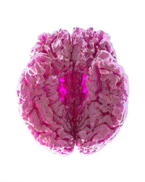
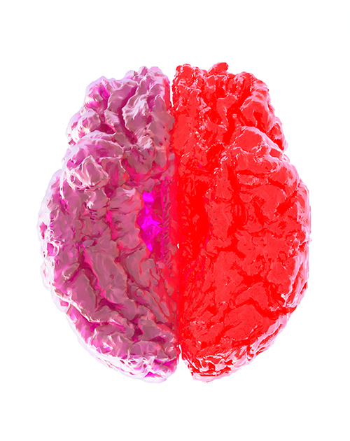
Hot Thoughts
MRI of Joe Borrellos Sliced and Renderd In CAD and manipulated in Photoshop.
The Friedman Brain Institute
One Gustave L. Levy Pl, New York, NY 10029
Contact: Veronica Szarejko (veronica.szarejko@mssm.edu)
#ArtoftheBrain